Consortium Lyon Saint-Etienne de Microscopie
The "Consortium Lyon Saint-Etienne de Microscopie" (CLYM, FED 4092) is a federative structure created in 1998 to have a pool of advanced microscopes, essentially electron microscopes, in order to finely characterize materials microstructures, from the micron scale to the atomic level. The application fields mainly belong to chemistry, physics, engineering and nanotechnologies. Among the machines hosted by CLYM, "environmental" microscopes allow in situ studies of materials behaviors under conditions mimicking their real use (operando microscopy). For that reason, CLYM is one of the platforms of the national network METSA.
In this context, its first mission is to manage microscopes that are partially or totally shared, so that its members can develop research activities related to materials microstructural characterization. The machines are located:
- At INSA Lyon, in premises close to those of the MATEIS laboratory,
- At CNRS, within the premises of the IRCELYON laboratory,
- At Jean-Monnet University in Saint-Étienne, within the premises of the Hubert Curien laboratory.
For CLYM partners, the microscopes can be used either in an autonomous manner after training, or in cooperation and under assistance of advanced users. For non-CLYM partners (laboratories or companies), experiments may be performed upon request (contact@clym.fr).
The expertise of CLYM in terms of materials characterization can also be found within the trainings delivered via CNRS Formations-Entreprises.
CLYM is one of the platforms of the labex iMUST, EUR Manutech-Sleight as well as Ingélyse and Institut de Chimie de Lyon federations.
ETEM 1
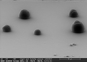
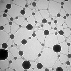 Condensation of water droplets at 1°C from a humid atmosphere (water vapor pressure 7 mbar introduced in the microscope in environmental mode). The droplets are formed on NaCl nanocrystals (dissolved) attached to the junctions of the filaments of a carbon lacey grid. ETEM 300 kV.
Credits: Eric Ehret, Francisco Cadete Santos Aires and Thierry Epicier (IRCELYON).
ETEM 2
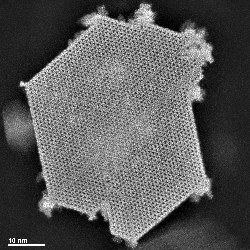 SSTEM HAADF image of a mixed oxide composed of two isomorphic phases doped with thallium. It is a catalyst used for the oxidation or ammoxidation of propane. The Mo7.8V1.2NbTl0.94O28.9 phase, called M1 phase, with orthorhombic structure, is the most active. The other major phase V2.2Mo6.6(Tlx)O24.2 has a trigonal structure.
Crédits : Mimoun AOUINE, Thi Minh NHA LE, Jean Marc MILLET (IRCELYON)
FIB
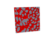 Silicon-rich phase grating present in an aluminum alloy, obtained with the CLYM double beam FIB/SEM microscope.
FIB volume: 5 x 5 x 0.325 μm³ - Voxel size target: (5nm)3
Photo taken as part of a study on the influence of the building platform temperature during selective laser melting of AlSi10Mg alloy.
Credits: Juan Guillermo Santos Macias, Aude Simar (Catholic University of Louvain), Thierry Douillard, Eric Maire (MATEIS).
AFM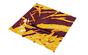
Map of the Young modulus distribution in a thermoplastic polyurethane (log scale). Image made using the PFQNM mode on the Bruker Dimension Icon atomic force microscope available within the CLYM. Field of view 5 µm x 5 µm.
Crédits : Florent Dalmas (MATEIS), David Albertini (INL).
ESEM
 Condensation of water droplets on aerosol particles. Image made in GSED mode on the QuattroS environmental scanning electron microscope of the CLYM. Image obtained during the development of an in situ nanotensiometer.
Credits: Clément Châtre (IRCELYON), Eric Ehret (IRCELYON), Philippe Steyer (MATEIS), Karine Masenelli-Varlot (MATEIS), Francisco Cadete Santos Aires (IRCELYON), Barbara Nozière (KTH, Sweden)
NEO-ARM
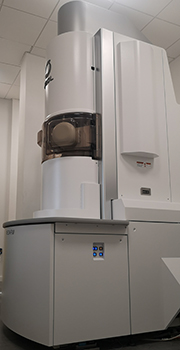
Picture of the JEOL NeoARM 200 FEG TEM transmission microscope, equipped with a cold source, a probe corrector, a 1sr EDS detector, and a Quantum Gatan GIF. The NeoARM is particularly well suited for STEM imaging and high-resolution STEM-EELS and EDS analytical characterization at 60, 80 and 200 kV.
This equipment was purchased by the Jean Monnet University, Saint-Etienne, thanks to CPER Manutech Europe 2015-2020 credits. It is shared within the Lyon Saint-Etienne Microscopy Consortium (CLYM).
The microscope is installed in the electron microscopy platform of the Hubert Curien laboratory, Site Manufacture, Saint-Etienne. The NeoARM Microscope team is made up of 5 ITAs and researchers from different laboratories: Stéphanie Reynaud (LABHC), Mimoun Aouine (IRCELYON) Matthieu Bugnet (MATEIS), Yaya Lefkir (LABHC), Anne-Magali Seydoux-Guillaume (LGLTPE).